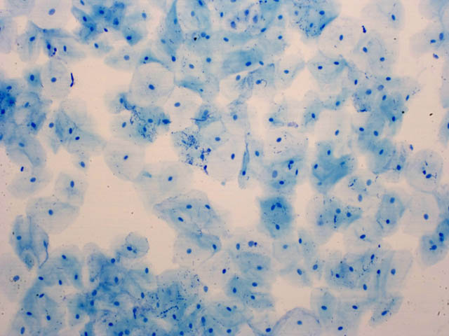
8. Glass with cell debris
On the edge of a glass that was on the table have been rests of cells of buccal epithelium. We are interested in knowing who they belong to, because they can give us important clues. We will look at the cells under the microscope and complete the CHEKING SHEET.
Practice: observation of cells of the buccal mucosa
Material
Oral mucosa samples
Cotton Sticks
Needle attached
Slides and coverslips
Dropper
Lighter
Microscope
Water
Methylene blue
Process
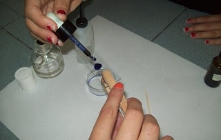
1. With the help of a cotton swab, we sampled the cells of the oral mucosa from three different partners.
2. With the needle take a small portion of the scraped and place the sample on a slide.
3. With the dropper, add a drop of water to the sample. With the same needle we mix it and extend it through the slide.
4. Let air dry (or heat a little on a lighter).
5. With the dropper, put one or two drops of methylene blue to the dry sample. Wait a minute and add a few drops of water to remove excess dye. 6. Cover the sample gently with the coverslip.
7. We observe the microscope.
Material
Oral mucosa samples
Cotton Sticks
Needle attached
Slides and coverslips
Dropper
Lighter
Microscope
Water
Methylene blue
Process
1. With the help of a cotton swab, we sampled the cells of the oral mucosa from three different partners.
2. With the needle take a small portion of the scraped and place the sample on a slide.
3. With the dropper, add a drop of water to the sample. With the same needle we mix it and extend it through the slide.
4. Let air dry (or heat a little on a lighter).
5. With the dropper, put one or two drops of methylene blue to the dry sample. Wait a minute and add a few drops of water to remove excess dye. 6. Cover the sample gently with the coverslip.
7. We observe the microscope.
|
|
Practice: Innoculate and streaking in Petri dishes.
Material:
Petri dish
Agar
Inoculation loop
Process:
1. Labelling of the Petri dishes;
2. Sterilisation of the inoculation loop
3. Take a sample of water conteined in a cover tuve.
4. Removal of the Petri dish cover;
5. Streaking;
6. Covering the Petri dish and puting in a fridge in a adequate temperature.
7. Covering the tube.
8. Observe the petri dish after 24 or 48 hours.
If you develope the previous experiments, you obtein 10 more points.

All right! We have completed all the cellular observations that are useful for the case.
We added the points obtained in this task and, if so far we add at least 30 points we put the award of SUPERBIOLOGISTS
We return to the beginning to continue solving the crime. Now we enter point 9
We added the points obtained in this task and, if so far we add at least 30 points we put the award of SUPERBIOLOGISTS
We return to the beginning to continue solving the crime. Now we enter point 9


