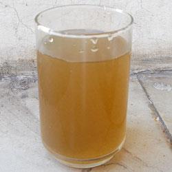
7. Bottle of mineral water
One of the evidence in Dr. John's office was a bottle of water lying on the floor. It is observed, by its dirty appearance, that someone has been able to introduce water contaminated with microorganisms in her. Professor Julius Martens was allergic to them. We will examine the sample and check the presence or absence of microorganisms. We will incorporate the results into the CHEKING SHEET

|
|
Practice: Microorganisms in water
Materials:
Bottles with water sample (infusion)
Microscope
Slides and coverslips
Filter paper
Dropper
Tweezers
Beaker
Neutral red dye
Microscope
Process:
1) We eject a little liquid from the bottom of the bottle and pour a drop in the center of several slides. In some we also put a few drops of neutral red dye.
2) Dry the excess liquid with a filter paper.
3) After covering them with coverslips we observe them under a microscope using small increases first and then moving to the highest magnification.
4) With the key we are classifying the microorganisms that we see. We draw or take pictures.
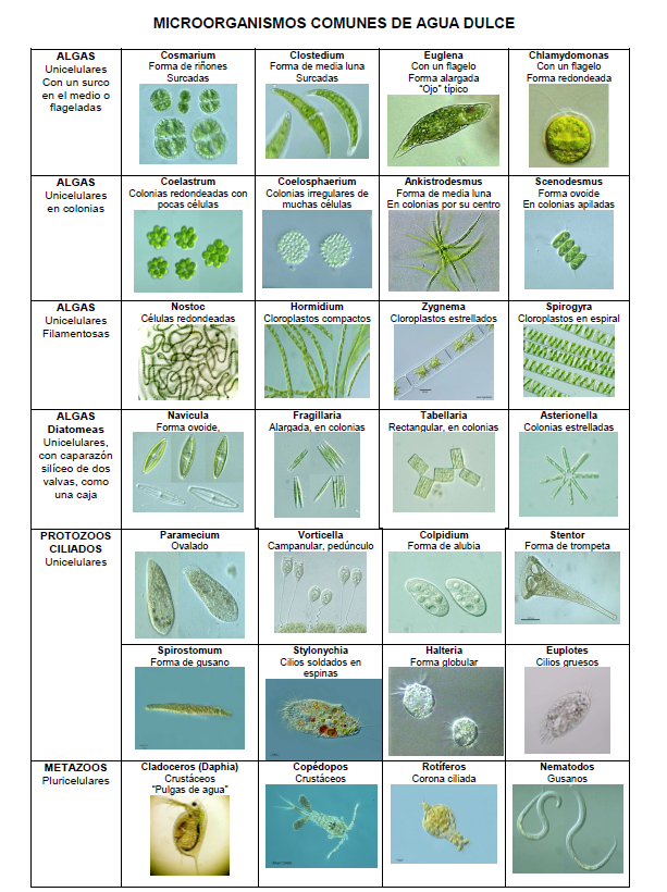
Among the most likely to find in a sample are:
1) Microscopic algae: In general they are very pigmented by chlorophyll, which allows their observation without need of staining. For example:
Diatoms. Unicellular, wrapped in a siliceous shell formed by two valves, like a box)
Demeidians. Unicellular with bright green chloroplasts and cells are divided into two halves by a furrow
Chlorophyceae: algae with chlorophyll, therefore have a green pigmentation. They can be unicellular or filamentous, these filaments being branched or not.
Cyanophyceae: algae with cyanine pigment, blue-green in color.
2) Protozoos: are organisms of very diverse forms, which move by means of cilia or flagella. Sometimes they appear in colonies, but they are always unicellular, never pluricellular. For example:
Ciliated: present cilia on its surface.
Flagellates: they have one or two flagella.
Rhizopods: protozoa that emit pseudopods for locomotion or feeding. Some families have exoskeleton (heliozoos and radiolarias).
3) Small metazoans of aquatic life. The most frequent are:
Rotiferous: small animals that present a continuous movement of their stomach masticador, well observable under the microscope.
Worms: very varied and large. The most frequent are nematodes, planarias and some annelids.
Crustaceans: the most abundant are copepods and daphnids.
Insects: in their larval forms.

Perfect!
We are already checking many data that bring us closer to figuring out who the killer is.
We return to the Beginning and continue with the 08, glass with remains of cells.
We are already checking many data that bring us closer to figuring out who the killer is.
We return to the Beginning and continue with the 08, glass with remains of cells.

