6. SANDWICH
|
In the office of Doctor John Carles, a sandwich has been found. A laboratory has performed a preliminary analysis of the sample and has sent us some photographs of its contents. Some are seen at first glance, such as cheese and ham:
|

|
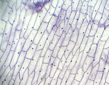
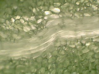
But others were crushed and cell samples were obtained like these:
|
|
We suspect one of them is onion and Andreu was allergic to it. We must check our suspicions in the cellular laboratory.
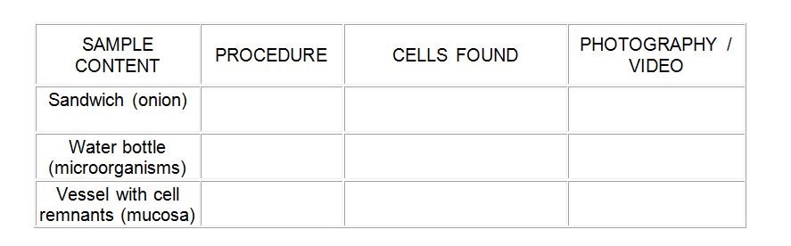
ACTIVITY 8:
Working in groups, we analyzed the samples in the laboratory. We will fill in the SHEET with the results and the methodology used, completing it with photographs or videos of the process. It will be evaluated with a corresponding rubric.
Firstly, you should read the manual process.
Working in groups, we analyzed the samples in the laboratory. We will fill in the SHEET with the results and the methodology used, completing it with photographs or videos of the process. It will be evaluated with a corresponding rubric.
Firstly, you should read the manual process.
Practice of onion epithelium
Materials:
An onion
Scalpel
Tweezers
Dropper
Methylene blue
Distilled water
Slides and coverslips
Microscope
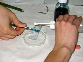
Process:
1 Cut the onion in half and, with the scalpel, remove a small piece of epidermis from the leaves of the bulb (transparent lamina) of not more than 1cm x 1cm.
2-With a tweezer we extract the tissue of the onion and place it extending well on a slide so that there are no air bubbles.
3-With a dropper we dyed with methylene blue and let it act for 5 minutes.
4-After the 5 minutes, the methylene blue has acted on the sample. It carefully removes the remains of the sample with distilled water. Then dry the sample in the air so that there are no traces of water.
5-We place the cover on the sample and take it under a microscope to observe the cell. With the macrometric and micrometric screws, we focus the cell until a clear image is obtained. The process always starts from the lens of smaller magnification. We observed in several magnifiers.
6- Draw our results in the following circles.
7- Check with the photographs of the samples and draw conclusions.
Materials:
An onion
Scalpel
Tweezers
Dropper
Methylene blue
Distilled water
Slides and coverslips
Microscope
Process:
1 Cut the onion in half and, with the scalpel, remove a small piece of epidermis from the leaves of the bulb (transparent lamina) of not more than 1cm x 1cm.
2-With a tweezer we extract the tissue of the onion and place it extending well on a slide so that there are no air bubbles.
3-With a dropper we dyed with methylene blue and let it act for 5 minutes.
4-After the 5 minutes, the methylene blue has acted on the sample. It carefully removes the remains of the sample with distilled water. Then dry the sample in the air so that there are no traces of water.
5-We place the cover on the sample and take it under a microscope to observe the cell. With the macrometric and micrometric screws, we focus the cell until a clear image is obtained. The process always starts from the lens of smaller magnification. We observed in several magnifiers.
6- Draw our results in the following circles.
7- Check with the photographs of the samples and draw conclusions.

|
|
|
Put 10 more points.
We go back to the beginning and continue to point 7 to examine the bottle that was on the floor |

|

